Foramen Spinosum
What is foramen spinosum?
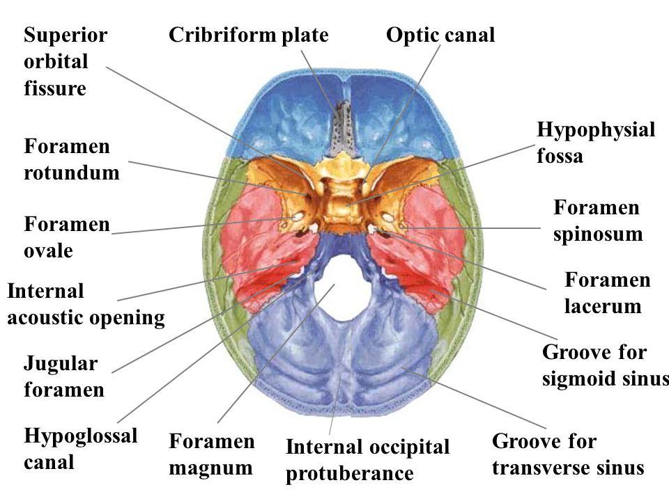
The foramen spinosum is an anatomical structure – foramen in the sphenoid bone, which is situated in the middle cranial fossa. The sphenoid bone has two foramina- foramen spinosum and foramen ovale, which is bigger.
Foramen spinosum was first discovered and described by anatomist Jakob Benignus Winslow in the 18th century. From Latin, the name foramen spinosum means “hole full of thorns”[1].
Foramen spinosum variations
Foramen spinosum can have several variations in position, size and its location. In rare cases, foramen spinosum is absent. This can happen if the middle meningeal artery enters the cranium through foramen ovale. Foremen spinosum can also be incomplete- this occurs in half of the population. Foramen ovale can also be duplicated, in case the middle meningeal artery is also duplicated. This occurs in less than 1% of the population [2].
![]()
Sphenoid bone
Sphenoid bone is one of the eight bones that make up the skull. The word sphenoid comes from Greek language and means “butterfly-shaped”. It lies in the middle cranial fossa and contributes to also making the lateral walls of the skull, floor and sides of both orbits.
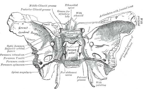
Sphenoid bone has four main parts: body, paired greater wings, lesser wings and two pterygoid processes. The body is the center of sphenoid bone and is almost cube-shaped. The body contains sphenoid sinuses which connect to ethmoid bone and open in the nasal cavity. The body also contains Sella turcica- a saddle-shaped depression in the bone and chiasmatic groove where the optic nerves partially cross.
The greater wing extends from the body and makes up three parts of the facial skeleton- floor of the middle cranial fossa, lateral wall of the scull and parts of the orbit. In the wing, there are also three foramina- foramen rotundum, foramen ovale and foramen spinosum. Through these openings, nerves and arteries pass in the cranial cavity. The lesser wing of sphenoidal bone separates the anterior and medial cranial fossa and form the lateral border of the optic canal [3].
![]()
Development
The foramen spinosum size in adults is around 2.56mm long and 2.1 mm wide. In newborns, this structure is about 2.25 mm long and 1.05 mm wide. Foramen spinosum develops its ring-shaped formation as early as eight months after birth, but it can also take shape as late as 8 years of age [4].
Functions
Foramen spinosum is an opening that allows several structures to pass through it. the following structures pass through foramen spinosum:
- Middle meningeal artery
- Middle meningeal vein
- Meningeal branch of mandibular nerve
Middle meningeal artery
Middle meningeal artery is a branch of maxillary artery. It passes through foramen spinosum and enters the middle cranial fossa. After entering the skull cavity, middle meningeal artery splits in two parts- superior tympanic branch and ganglionic branch and divides into anterior and posterior division. Anterior division goes to the anterior side of the sphenoid bone and gives its terminal branches in the upper parietal bone. The posterior division runs to the posterior side over temporal bone and gives its terminal branches in lower parietal bone.
The branches of this artery run above dura mater, which protects the brain tissue. Damage to the artery can cause epidural bleeding- accumulation of blood over dura mater. Such damage can be caused if foramen spinosum or temporal bone is traumatized [5].
Middle meningeal vein
Middle meningeal vein runs parallel to the middle meningeal artery. It emerges from the pterygoid plexus and joins the superficial temporal vein, subsequently becoming retromandibular vein. It later drains into the subclavian vein.
Epidural hematomas from a rupture of vein are seen in 40% of cases, the 60% being from rupture of artery. Venous bleeding from middle meningeal vein is usually caused by skull fractures and is mostly a problem in children [6].
Foramen spinosum is also an anatomical landmark during surgery. It helps to reveal positions of the mandibular nerve and trigeminal ganglion, as well as foramen ovale and foramen rotundum. It is also relevant, when homeostasis has to be achieved during surgery [7].
References
- Location: http://link.springer.com/article/10.1007%2Fs10143-008-0152-6
- Variations: http://www.anatomyatlases.org/AnatomicVariants/SkeletalSystem/Text/SphenoidBone.shtml
- Sphenoid bone: http://teachmeanatomy.info/head/osteology/sphenoid-bone/
- Development: https://www.ncbi.nlm.nih.gov/pubmed/3610040
- Middle meningeal artery: http://www.healthline.com/human-body-maps/middle-meningeal-artery
- Middle meningeal vein: http://www.healthline.com/human-body-maps/middle-meningeal-vein
- Clinical significance: https://www.ncbi.nlm.nih.gov/pmc/articles/PMC3836884/


