Asystole
What Is Asystole?
Asystole refers to an abnormal cardiac rhythm as indicated by an electrocardiogram (ECG) machine. If this is identified on a patient, there is a treatment protocol to follow return the heart rhythm to normal [1, 2, 3].
The definition of asystole is a type of a cardiac arrest rhythm that has no distinct electrical activity seen on the ECG machine. The waves and complexes typically found in a normal heart beat are not present [2].
It indicates that the heart is not functioning anymore and this life-threatening condition needs immediate attention to save the patient [1, 2, 3]. An asystole is a condition used by the physician to verify the clinical death of a patient [2]. Figure 1 shows the difference between a normal ECG reading and that of asystole.
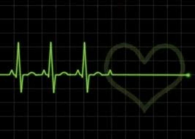
Figure 1- Normal ECG and Asystole
The sinoatrial node (SA node) of the heart serves as the pacemaker and is responsible for the electrical impulses that cause the cardiac muscles in the atrium to contract.
The atrioventricular node (AV node) send the electrical impulses in the ventricles. Problems in the SA node can cause arrhythmias or abnormal heart rhythms. An arrhythmia can cause the heart to beat too slow, too fast or stop it from beating again [2].
Types
There are 2 known types of asystole, primary asystole and secondary asystole [2].
Primary asystole
In a primary asystole, the SA node is unable to generate an electrical impulse to allow depolarization of the ventricles. This occurs when there is a degeneration or ischemia in the SA node or the AV node. Another arrhythmia usually precedes a primary asystole which causes a block-arrest in the SA node or a complete heart block [1, 2, 3].
Secondary asystole
This type of asystole happens when outside factors cause the heart to fail to generate the electrical impulse. This usually occurs if the ventricular tachycardia is untreated and after unsuccessful defibrillation attempts [1, 2, 3].
Causes
The development of asystole is associated with some factors. The causes of primary asystole are different from the ones of secondary asystole [1, 2, 3, 4].
Primary asystole
Tissue ischemia is one of the main causes of primary asystole. It prevents the electrical impulses in the SA node to be generated and passed on to other cardiac cells. Occlusion of the coronary arteries also causes ischemia in the SA and AV node. Indirect lightning strike or electrocution can lead to asystole. A local tumor, cardiac trauma, or a failure of a cardiac pacemaker to work properly can lead to this type of asystole [1, 2, 3,4].
Secondary asystole
Medical conditions that may cause secondary asystole include stroke, hyperkalemia or increased potassium levels in the blood, myocardial infarction, pulmonary embolism, ventricular tachycardia, overdoses form hypnotic drugs or narcotics, near drowning and suffocation. Hypothermia may also lead to a secondary asystole but the body is able to tolerate it for a longer period of time [1, 2, 3, 4].
Symptoms
The diagnosis of asystole is based on the recognition of a heart rhythm that has no discernible electrical activity. The electrical activity of the heart is assessed with the use of an ECG machine. If bradycardia or decreased heart rate occurred before the asystole, the patient may present with syncope or light-headedness.
If the patient is at a cardiac standstill for more than several seconds, the patient may become unconscious and unresponsive. There may be no detectable heart sounds and the peripheral pulses will not be palpable.
If the condition is not managed in 15 minutes or more, the deprivation of oxygen in the brain may lead to brain death [1, 2, 3, 4].
Treatment and Management
The initial management of patients who present with asystole in the emergency department is to provide oxygenation, ventilation, and circulation to the patient. This is achieved by performing an endotracheal intubation and cardiopulmonary resuscitation (CPR).
Pharmacologic agents are also given to the patient at this point. The resuscitation will continue until the patient re-establishes a normal cardiac rhythm. CPR may be stopped if rigor mortis have already set in the patient [1, 2, 3, 4].
Electrical defibrillation is not performed in patients in asystole because it will remove any chance of the patient to recover a cardiac rhythm. The outcome of asystole that occurs after electrical defibrillation is worse than in patients whose first documented rhythm was asystole [1, 2, 3, 4].
If the patient survives the cardiopulmonary arrest, they may be transferred to an intensive care unit for further treatment and evaluation. There is evidence that suggests a better neurologic outcome in a patient who are cooled to around 32-34°C during the first 24 hours after the arrest [1, 2, 3, 4].
Prevention
The development of asystole in patients who have presented with a heart block may be prevented through the use of a permanent pacemaker. They must follow the precautions regarding the pacemaker to ensure that it is functioning properly. Prevention of a secondary asystole can be done by managing the patients underlying medical condition [1, 2, 3].
References
- Shah, S. N. (2015, December 26). Asystole. Retrieved from Medscape: http://emedicine.medscape.com
- Practical Clinical Skills. (2016). Asystole. Retrieved from Practical Clinical Skills: https://www.practicalclinicalskills.com/asystole
- Study.com. (2016). Asystole: Definition, Causes & Treatment. Retrieved from Study.com: http://study.com/academy/lesson/asystole-definition-causes-treatment.html
- Carey, M. G., Al-Zaiti, S. S., & Pelter, M. M. (2010). Asystole. American Journal of Critical Care, 84-85.


